
Figure 3.
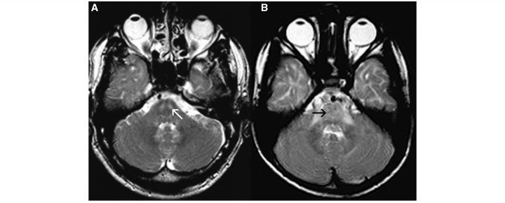
Axial T2-weighted images of two male patients with PMLD (age 39 years in A, age 6 years in B) at the level of the pons show T2 hyperintensity, either in the pyramidal tracts alone (white arrow, A) or in the entire pons (black arrow, B).
Official websites use .gov
A
.gov website belongs to an official
government organization in the United States.
Secure .gov websites use HTTPS
A lock (
) or https:// means you've safely
connected to the .gov website. Share sensitive
information only on official, secure websites.
Axial T2-weighted images of two male patients with PMLD (age 39 years in A, age 6 years in B) at the level of the pons show T2 hyperintensity, either in the pyramidal tracts alone (white arrow, A) or in the entire pons (black arrow, B).