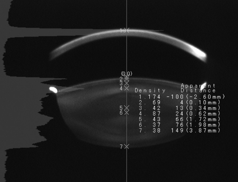
Fig. 1.
Scheimpflug slit image obtained using an anterior eye segment analysis system Digitalized anterior eye segment image is demonstrated at a mid-sagittal section, the front of the cornea is at the top of the image. The seven numbers depicted on the axis sequentially indicate the cornea (1), anterior capsule (2), most transparent layer of the anterior superficial cortex (3), anterior adult nucleus (4), anterior fetal nucleus (5), central clear zone (6) and posterior subcapsular region (7). The peak light scattering intensity for each segment is demonstrated to the right of each respective layer.

