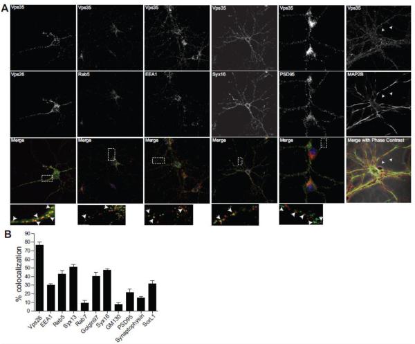
Figure 1. Vps35 localizes on endosomes and trans-Golgi Network (TGN) throughout the neuron.
A, 14 day old mouse primary hippocampal neurons from postnatal day 0 mice were fixed and stained for endogenous Vps35 (red) and the indicated organelle or synaptic markers (green) using antibodies, and imaged using confocal microscopy (Zeiss LSM 700). Arrows in the inset show regions of colocalization. B, Colocalization index for the indicated stainings. Values denote Means ± SEM (n ≥ 25 fields). Scale bars are 10 μm in all panels.

