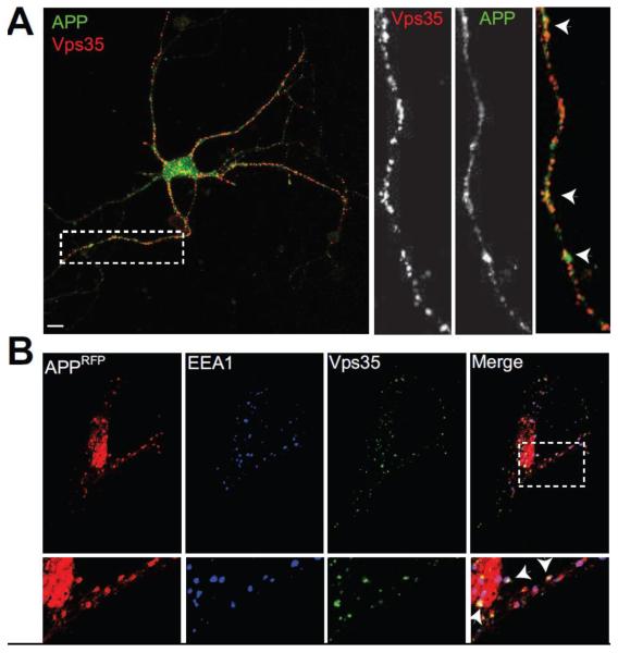
Figure 2. Vps35 colocalizes with Amyloid Precursor Protein.
A, Hippocampal neurons (14 DIV) were fixed and stained for endogenous Vps35 (red) and the Amyloid Precursor Protein (APP) (green). Neurons were imaged as in Fig. 1. Inset shows colocalization between APP and Vps35 occurring also in neuronal processes. B, HeLa cells were transfected with APP-mRFP (red), and stained for endogenous Vps35 (green) and the early endosomal protein EEA1 (blue), 24 hours post transfection. Merge shows extensive colocalization between Vps35 and APP occurring on early endosomes. N=15 fields from at least three independent experiments. Scale bars are 10 μm.

