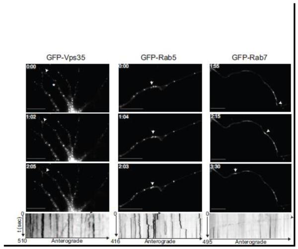
Figure 5. Vps35 exhibits short-range movement in hippocampal neurons.
Hippocampal neurons (9 DIV) were transfected with GFP-Vps35 (left panel), GFP-Rab5 (middle panel) or GFP-Rab7 (right panel), and imaged 24-48 hours post transfection. Time-lapse images were collected using a GFP filter every 5 seconds for the indicated times. Representative time-lapse images show Vps35 (left) positive puncta exhibiting a short-range jitter-like movement similar to Rab5 (middle), whereas Rab7 undergoes long-range movement in neuronal processes. White arrows in the images correspond to the black arrows in the kymographs below, highlighting either the local (for Vps35 and Rab5) or long-range movement (for Rab7). Kymographs were generated from the time-lapse movies using MetaMorph software. The star in Vps35 panel depicts the neuronal process for which the kymograph is shown below. N=3 for Rab5 and Rab7. N>3 for Vps35. Scale bars are 10 μm in all panels.

