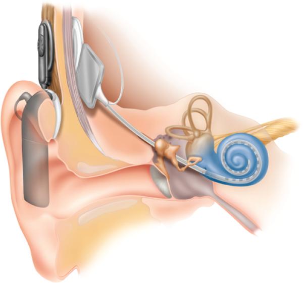
Fig. 2.
Illustration of the cochlear implant. The device consists of an external microphone and processor, which connects to an internalized receiver and electrode array through the scalp. The linear electrode is placed within the cochlea. Illustration provided courtesy of Cochlear™ Americas, ©2009 Cochlear Americas.

