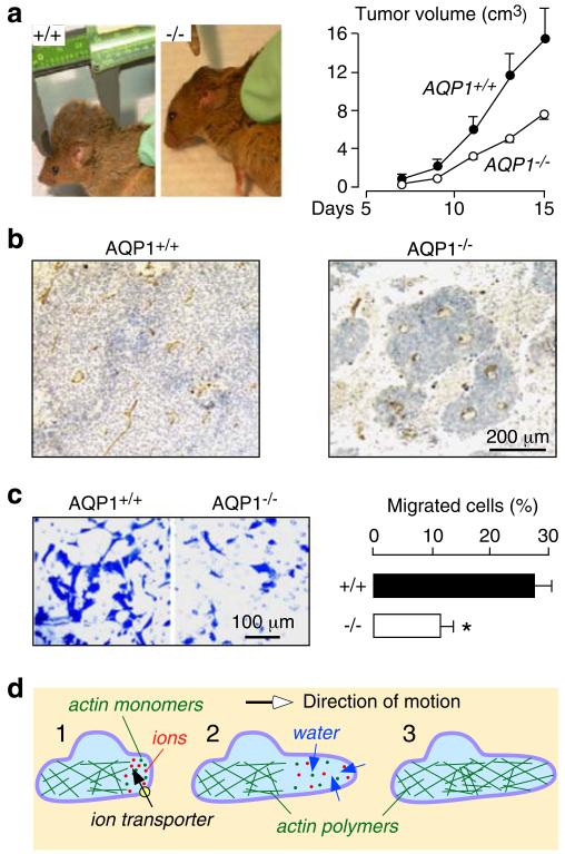
Fig. 1.
Impaired angiogenesis and endothelial cell migration in AQP1 knockout mice. a Tumor in wild-type vs AQP1 null mouse, 2 weeks after subcutaneous injection of 106 B16F10 melanoma cells (left). Tumor growth data (10 mice per group, P<0.001) (right). b Tumor tissue stained with the endothelial marker isolectin-B4 (brown). c Migration of aortic endothelial cells for 6 h towards 10% serum shown in a transwell assay (left). Picture shows migrated wild-type and AQP1 null endothelial cells (stained with Coomassie blue) after scraping. Data summary (S.E., n=16–20, asterisk P<0.001) (right). d Proposed mechanism of AQP-facilitated endothelial cell migration: 1 Actin depolymerization and ion movements increase osmolality at the anterior end of the cell. 2 Water entry increases local hydrostatic pressure, producing cell membrane expansion to form a protrusion. AQP polarizes to the leading edge of the cell membrane facilitating water entry into the cell. 3 Actin repolymerizes stabilizing the protrusion. Adapted from [14, 17]

