
Figure 1. Neuroimaging.
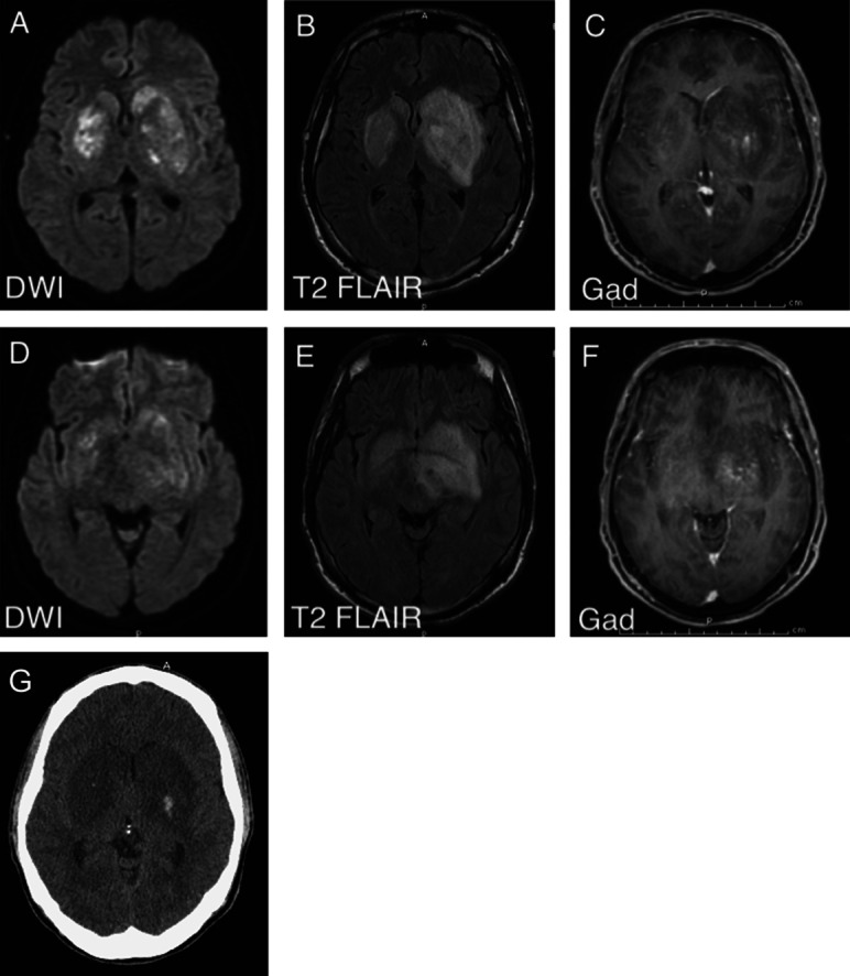
(A) MRI diffusion-weighted imaging (DWI) shows restricted diffusion in the lateral basal ganglia. (B) T2 fluid-attenuated inversion recovery (FLAIR) shows vasogenic and cytotoxic edema in the bilateral basal ganglia. (C) T1 postcontrast image shows minimal enhancement in the left basal ganglia. (D, E, F) Corresponding DWI, T2 FLAIR, and T1 postcontrast sequences at the level of the midbrain. (G) CT scan showing hemorrhagic transformation of the bilateral basal ganglia lesions on hospital admission day 3.