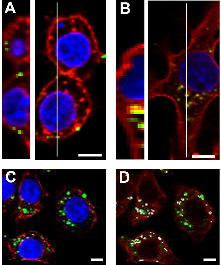
Figure 2. CPMV is bound and internalized by RAW264.7 macrophages and accumulates in late endosomes.
(A) CPMV-AF647 was incubated with RAW264.7 macrophages for one hour at 4 °C with 1 × 10⁶ viruses/cell. (B) CPMV-AF647 was incubated with RAW264.7 macrophages for four hours at 37 °C with 1 × 10⁶ viruses/cell. Slices through cells were imaged by confocal microscopy and multiple slices in a Z-series at the white line in left images and were compiled using ImageJ to make images on right. Bar, 5 µm. (C–D) RAW264.7 macrophages were incubated with CPMV-AF647 for four hours at 37 °C. Confocal microscopy imaging revealed colocalization (white, D) of CPMV fluorescence (green) and fluorescent antibody staining of LAMP-2 (red).

