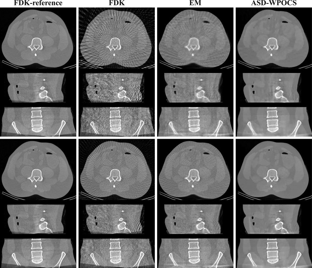
Figure 11.
Images reconstructed from 72-view (rows 1–3) and 120-view (rows 4–6) pelvis-phantom-data sets by use of the FDK, EM, and ASD-WPOCS algorithms, within a transverse slice at z = 0 cm (rows 1 and 4), coronal slice at x = 1.0 cm (rows 2 and 5), and sagittal slice at y = −1.0 cm (rows 3 and 6). Display window: [0, 0.35] cm−1.

