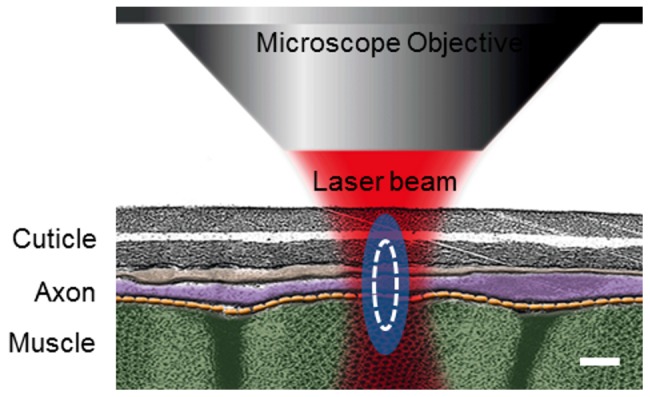
Figure 1. TEM illustration of the anatomical region of C. elegans where the laser axotomy is performed.

. Body wall muscles are shown in green, axons in purple and cuticle in gray. Blue ellipsoid is the estimated full-width-at-half-maximum of the beam's point spread function. White line represents the limit of the maximum plasma density generated at the focal spot. Scale bar 300 nm.
