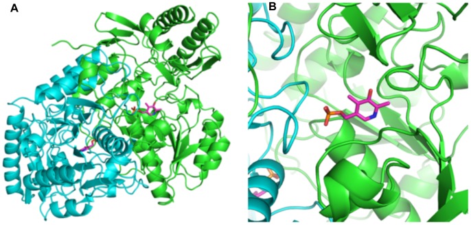
Figure 4. The native AstC structure with PLP.
Figures 4A and 4B show the native dimer structure as a ribbon Cα trace with the PLP cofactor shown in a magenta colored stick representation. Figure 4B is a zoomed image showing how the loop of residues 274–284 comes in from the neighboring protomer to contact the PLP cofactor and form part of the active site of protomer A.

