
Figure 3. AMD3465 inactivates CXCR4 in 4T1 tumors and slows tumor progression.
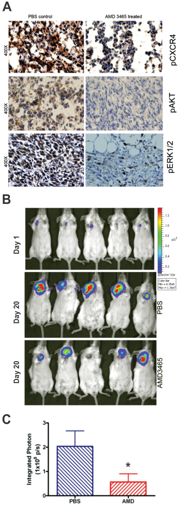
A, Tumor-bearing mice were injected with AMD3465 a single subcutaneous dose of 2.5 mg/kg. The tumor tissue was collected 1 h after treatment and sectioning was carried out. Immunohistochemical staining of pCXCR4, pAKT and pERK1/2 positive tumor cells can be seen in PBS controls compared to AMD3465 treated tumor sections. The slides were analyzed with an Olympus BX 41 microscope equipped with a digital camera (Olympus DP70). B, The top panel illustrates the BLI of 5 representative mice 1 d after injection of the 4T1 cells. The middle panel shows representative BLI in 5 mice treated with PBS 20 d after tumor injection, and the lower panel displays imaging of the 4T1 tumor masses following a similar exposure to AMD3465 (please see the Methods section for details). C, A bar graph representation of the end-point integrated photon (photons/cm2/sec) data collected in the experiment described in B. The tumor size was measured by BLI between control mice (n = 5) and AMD3465-treated mice (n = 10) and are expressed as the mean value ± SD (*p<0.05).