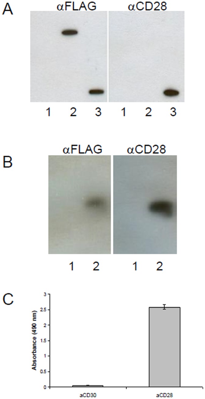
Figure 1. Analysis of recombinant CD28 expression in HEK cells.

(A) Coimmunoprecipitation. Recombinant HEK cells were lysed with lysis buffer, and 200–500 µl of cell lysate was incubated with rabbit αFLAG antibody at 4°C for 2 hours, then 20 µl of protein A agarose slurry (GE Healthcare) was added for another 2 hours. The beads were washed three times with at least 10 volumes of lysis buffer before resolving by SDS-PAGE. Detection was done either with mouse αFLAG or mouse αCD28. As control HEK293-SLP2-FLAG was used. 1: HEK293 lysate, 2: HEK293-CD28-FLAG lysate, HEK293-SLP2-FLAG lysate. (B) Westernblot. Cells were lysed and analysed by immunoblot using αFLAG or αCD28 antibodies. 1: HEK293 lysate, 2: HEK293-CD28-FLAG lysate (C) Elisa. Recombinant CD28 is recognized by a commercial αCD28 mAb. HEK293-CD28-FLAG lysate is coated on NUNC maxisorp via FLAG-tag. Detection was done with 1: αCD30 or 2: αCD28.
