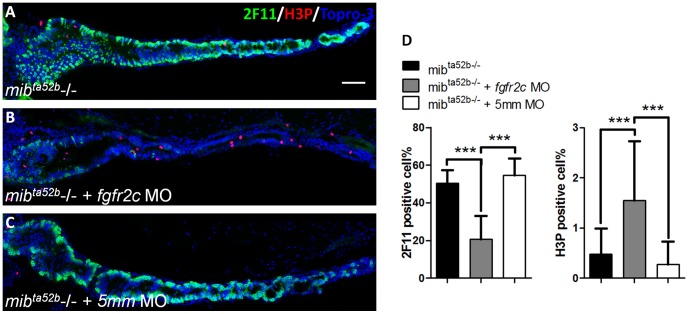
Figure 8. The secretory cell differentiation of mibta52b mutants after injection with fgfr2c morpholino.
The 2F11 (green) and H3P (red) antibodies were used to label the secretory and proliferating cells respectively, in (A) mibta52b mutant, (B) mutant injected with fgfr2c morpholino, and (C) mutant injected with fgfr2c-5 mm morpholino at 5 dpf. Topro-3 was used for nuclear counter staining (blue). (D) The bar charts show the percentages of secretory and proliferating cells. Error bars indicate SD. Scale bar = 50 µm.

