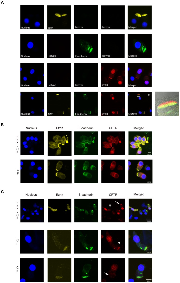
Figure 1. Immunolocalization of CFTR in nasal epithelial cells by confocal microscopy.
Nasal cells were collected on ice, enriched for single cells, fixed and stained for Ezrin (yellow), E-cadherin (green), and CFTR (red). Nuclei are indicated by DAPI (blue). A. Specificity of the antibodies was assessed by comparison with corresponding isotype controls. Differential interference contrast image was included in the last merged picture. B. Representative examples of non-CF and CF columnar epithelial cells with similar Ezrin and E-cadherin staining. C. Representative examples of variability in apical CFTR expression within columnar epithelial cells from one individual. Upper panel; cells obtained from a non-CF individual. Lower two panels; cells obtained from an individual with CF (F508del/F508del). White arrow points to apical membranes with or without CFTR.

