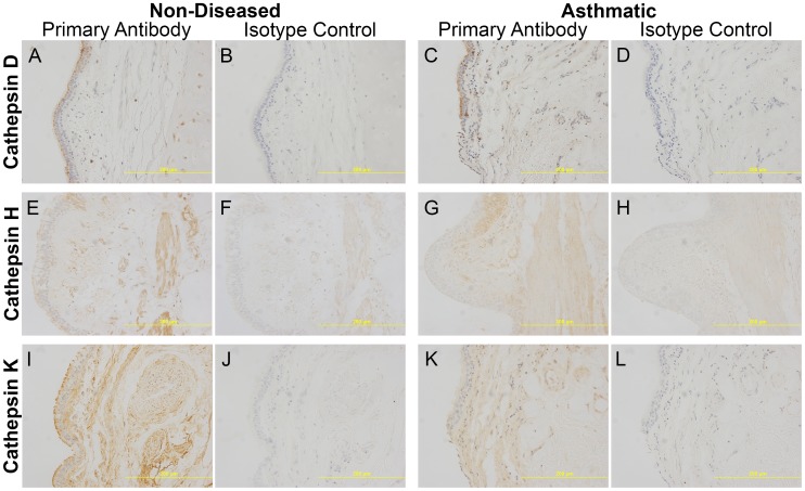
Figure 3. Representative images (20x magnification) of airway sections stained for cathepsins.
Immunostaining of cathepsins and corresponding isotype controls for cathepsins D (A–D), H (E–H) and K (I–L) from non-diseased and asthmatic sections. Specific staining was detected using a chemical chromophore DAB (brown) and cell nucleus was counterstained with haematoxylin (blue). Abbreviations DAB = 3,3′-diaminobenzidine.

