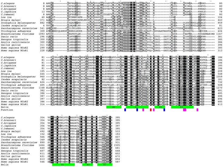
Figure 1. Comparison of N-terminal regions of GEI-8-related proteins to NCoR/SMRT.
Sequence alignment of GEI-8 nematode orthologues with their nearest Metazoa/Fungi homologues, both human orthologues NCoR1 and SMRT (NCoR2) are shown. Green bars indicate the position of the alpha-helices in the structure of the upstream DAD domain of human SMRT and homology predicted positions in the second SANT domain. Residues indispensable for regulating HDAC interactions and function are highlighted in blue (needed for the structural integrity), magenta (interaction with HDAC) and red (activation of HDAC). Only the N-terminal part of the sequences is shown. The identical and similar residues are highlighted by different intensity of shading. Sequence identifiers: C. elegans: GEI8_CAEEL, C. brenneri: CN15693, C. briggsae: A8X8F0_CAEBR, C. remanei: RP40355, C. japonica: JA23925 ABLE03010463.1 ABLE03032768.1 ABLE03032771.1 ABLE03032769.1 ABLE03032772.1, Loa loa: E1FVE0_LOALO, Brugia malayi: A8NSC3_BRUMA, Ixodes scapularis: B7PZ26_IXOSC, Saccharomyces cerevisiae: SNT1_YEAST, Drosophila melanogaster: Q9VYK0_DROME, Trichoplax adhaerens: B3SAN1_TRIAD, Branchiostoma floridae: C3XV35_BRAFL, Danio rerio: A8B6H7_DANRE, Xenopus tropicalis: NCOR1_XENTR AAMC01044136.1, Anolis carolinensis: ANOCA15679 2 ENSACAP00000014806; ENSACAT00000015107, Gallus gallus: UPI0000E813A6, Homo sapiens: NCOR2_HUMAN (NCoR2), Homo sapiens: NCOR1_HUMAN (NCoR1). PDB structure 1XC5 was used to determine the position of the helices.

