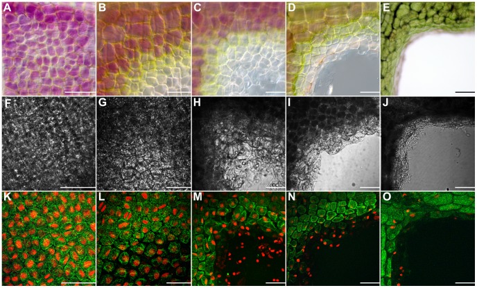
Figure 2. Rearrangement of the actin cytoskeleton during leaf morphogenesis over five stages of leaf development in the lace plant.
Each image depicts a piece of a single corner of an areole (A–E). Coloured differential interference contrast (DIC) images of (A) pre perforation (B) window (C) perforation formation (D) perforation expansion and (E) mature stage leaf areoles. (F–J) Black and white DIC images of (F) pre perforation (G) window (H) perforation formation (I) perforation expansion and (J) mature stage leaves. (K–O) Fluorescent images of Alexa Fluor 488 phalloidin (green) stained areoles counterstained with propidium iodide (PI; red) of (K) pre perforation (L) window (M) perforation formation (N) perforation expansion and (O) mature stage leaves. Please note that images A-E do not correspond with fluorescent images F-O. These images are representative micrographs to illustrate the five stages of leaf development. DIC images F-J are corresponding to K-O. There is a consistent gradient of actin microfilament staining over the pre perforation areole; conversely there is variation (bundling followed by breakdown) in actin microfilament dynamics over the gradient of PCD (NPCD-LPCD) found within the single areole of the window stage leaf. Note the complete degradation and disappearance of actin microfilament staining as the perforation becomes larger, from perforation formation to the mature stage of leaf development. Scale bars = 70 µm.

