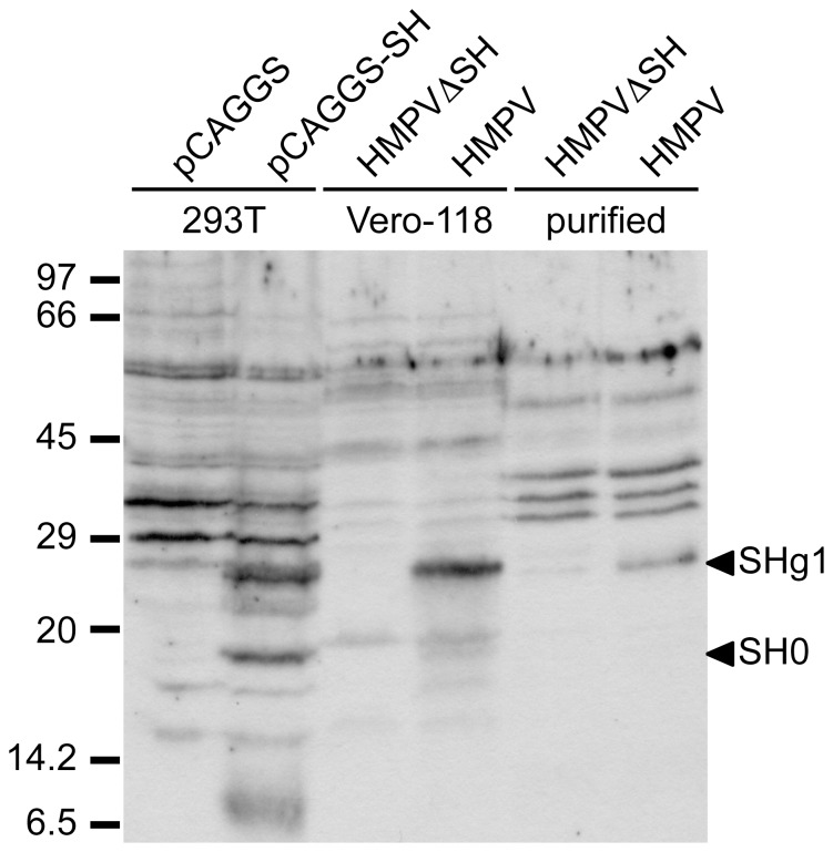
Figure 3. Western blot analysis of the SH protein.
293T cells transfected with pCAGGS (lane 1), pCAGGS-SH (lane 2), Vero-118 cells infected with HMPV (lane 3) or HMPV ΔSH (lanes 4) and purified virions of HMPVΔSH (lane 5) and HMPV (lane 6) were analyzed on 12.5% polyacryamide gels. The SH protein was detected using rabbit serum raised against a mixture of petides representing aa 2–16 and 95–110 of the SH protein.

