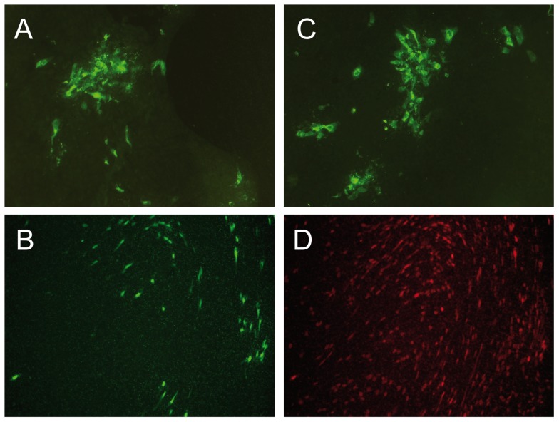
Figure 4. HPBEC cultured at air-liquid interphase were inoculated with wild type HMPV (a, b and d) or HMPVΔSH (c) at a MOI of 4.
Six days after inoculation, infected cells were visualized by immunostaining with HMPV specific polyclonal anti-serum (a and c). The HMPV-infected cells from panel b and d are the same field of cells, double stained for HMPV infected cells (b) and ciliated cells by staining with anti ß-tubulin antibodies (d).

