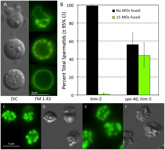
Figure 9. The status of the membranous organelles (MOs) in sperm.
A) When labeled with the membrane dye FM® 1–43 (Life Technologies™), fused MOs are visible as bright spots just inside the cell membrane, but unfused MOs are not labeled. Here, the sperm are shown in both DIC illumination and epifluorescence illumination. Top panel: a spermatozoon with an obvious pseudopod and the bright foci in the cell body indicating fused MOs. Middle panel: an abnormal spermatid with fused MOs. Bottom panel: a normal spermatid with no fused MOs. B) The percentage of normal spermatids and those that had at least a single obvious fused MO from him-5 and spe-46; him-5 male worms. Error bars represent SEM. In C-F, the sperm have been fixed, permeabilized, and exposed to an Alexa Fluor® 594 conjugate of wheat germ agglutinin (WGA; Life Technologies™), which labels all MOs, including those that have not fused with the cell membrane (original red fluorescence false colored green). C) Sperm from a spe-46(hc197); him-5(e1490) male. D) The same cells as is C, visualized in DIC optics. E) Sperm from a him-5(e1490) male labeled as in C. F) DIC image of the cells in E.

