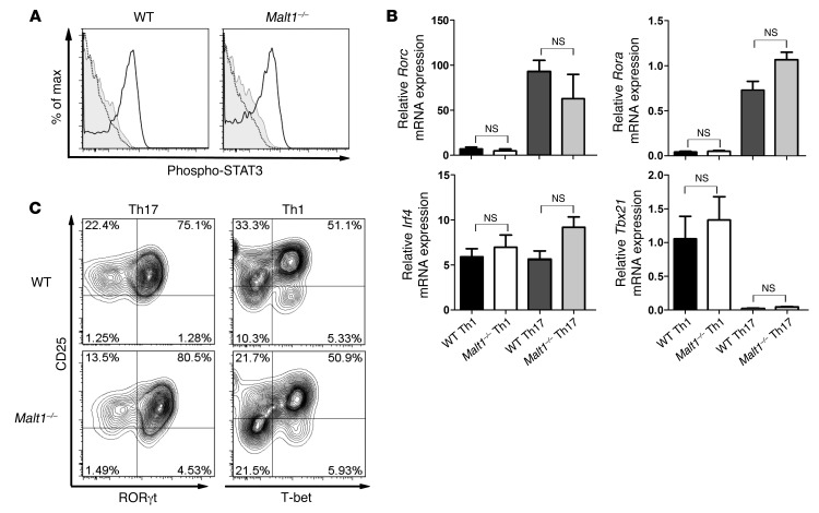
Figure 5. MALT1 does not interfere with expression of Th17-associated transcription factors.
(A) Malt1–/– and WT naive CD4+CD62L+ T cells were treated with IL-6 for 30 minutes (black line), and phospho-STAT3 levels were assessed by intracellular staining and flow cytometry. Gray line, isotype control; dotted line, without IL-6. Data are gated on live CD4+ T cells and representative of 3 independent experiments. (B) Levels of Th17 cell lineage–specific transcription factors were determined in Malt1–/– and WT Th1 and Th17 cells. Th cell differentiation was induced as described in Methods. After 16 hours of priming, mRNA levels of the indicated transcription factors were determined by RT-PCR. Results are expressed as the mRNA level normalized to Hprt and relative to that in naive WT Th cells. Data are mean ± SEM of 3 independent experiments. (C) In vitro differentiation of WT and Malt1–/– Th1 and Th17 cells was induced as in Figure 2. Expression of the indicated transcription factors was measured by intracellular flow cytometry. Data are gated on live CD4+ T cells and are representative of 4 independent experiments.

