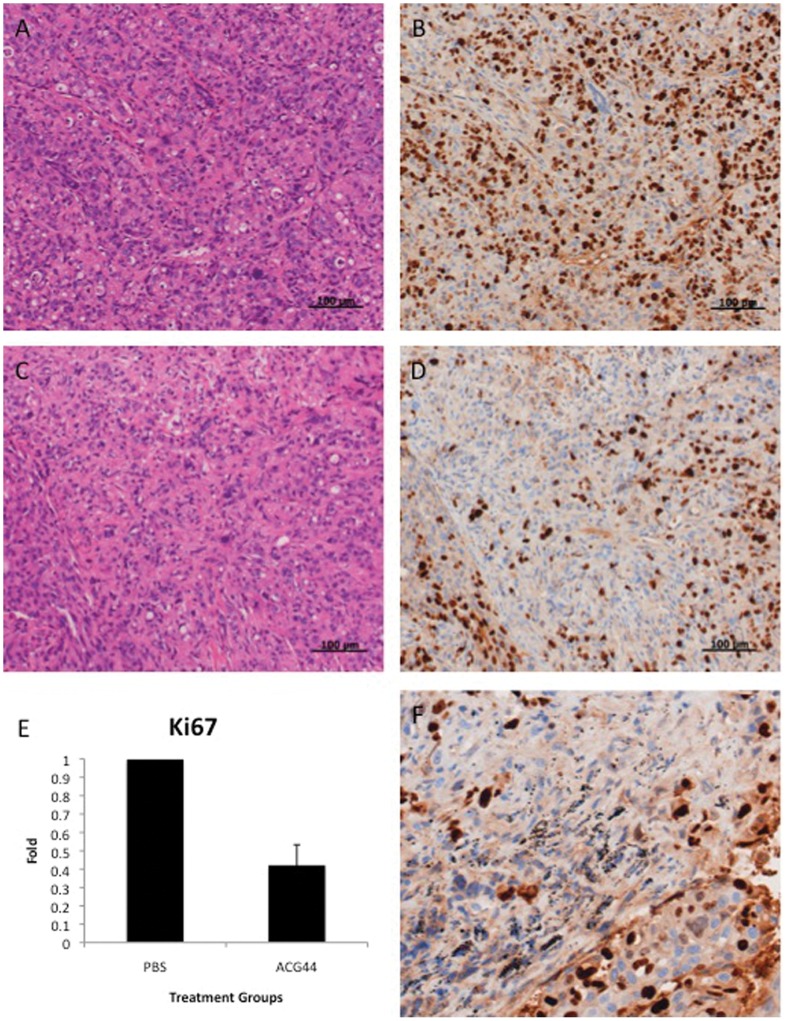
Figure 4. Immunohistochemistry Analysis of Tumors from the PBS and ACG44 groups.
Figure 4A and 4B show representative images of H&E and Ki-67 stained tumor tissues, respectively, from the PBS treated group whereas Figure 4C and 4D show images of H&E and Ki-67 staining of tumor tissue from the ACG44 treated group. All images were taken with 20× magnification. Figure 4E is quantification of the Ki-67 positive proliferative nuclei shown in Figures 4B and 4D. Figure 4F is a tumor image of Ki-67 staining from the ACG44 treated group, taken at 100 X to show gold accumulation (black spots) at a high magnification.

