
Fig. 2.
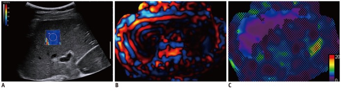
Shear wave elastography (SWE) and magnetic resonance elastography (MRE) images in of 39-year-old liver donor.
Liver stiffness map on SWE (A) was color-coded with blue, which suggested lower liver stiffness value. Liver stiffness value on SWE was 2.9 kPa. On MRE wave image (B) and elastogram (C), liver showed narrow wavelength over liver, as well as lower stiffness value, which was color-coded with violet and blue.