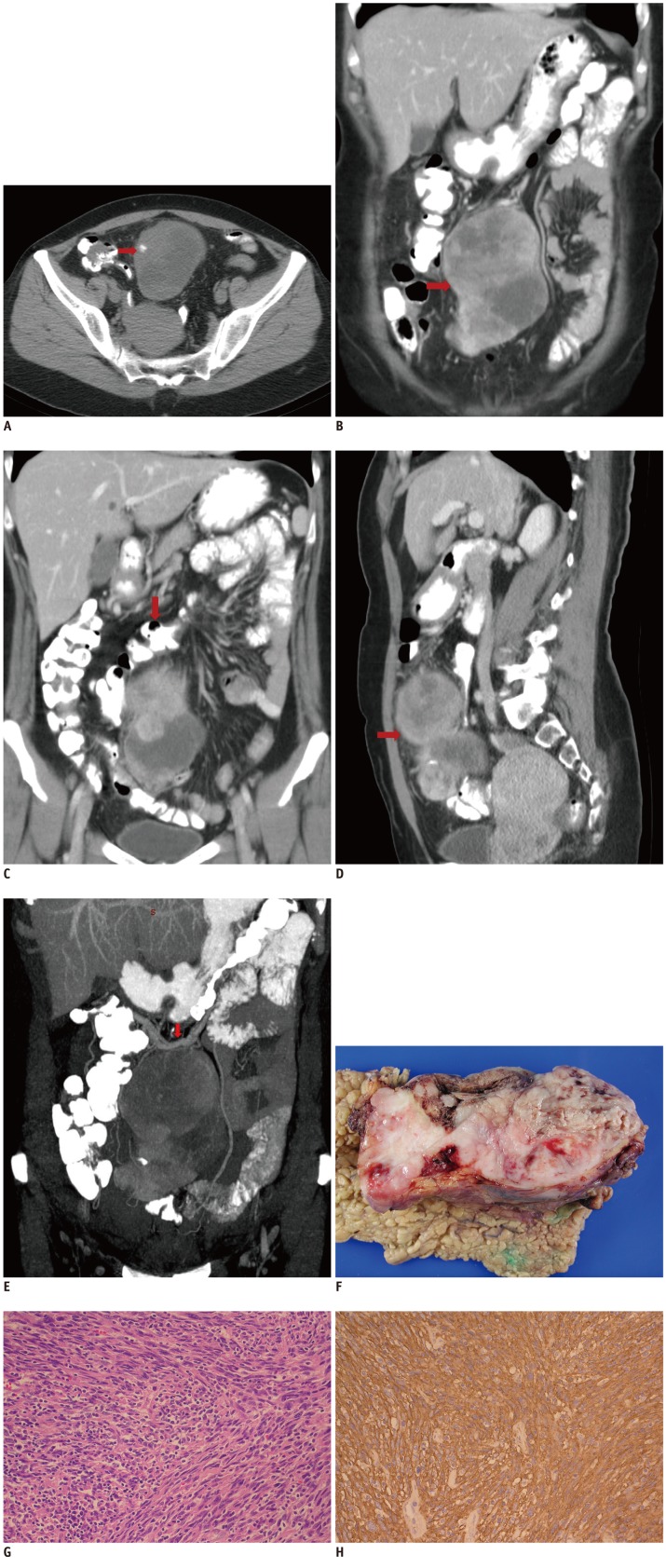
Fig. 1.
Follicular dendritic cell sarcoma in 47-year-old woman.
A. Precontrast CT scan in axial plane displays coarse chunk-like calcification within tumor. B. Post-contrast CT scan of whole abdomen in coronal plane reveals heterogeneously enhanced soft-tissue mass below stomach (arrow). C. Coronal postcontrast CT scan shows relationship between soft-tissue mass and transverse colon (arrow). D. Sagittal postcontrast CT scan shows tumor mass draped below transverse colon (arrow). E. Coronal maximum intensity projection image shows engorged right gastroepiploic vessels (arrow) with branches to tumor. F. Gross specimen of tumor shows well encapsulated, fleshy mass from greater omentum. G. Tumor is mainly composed of spindle cells in storiform and whorl arrangement. Varying numbers of lymphocytes admixed in tumor tissue are shown (H&E, original × 200). H. Tumor cells are diffusely positive for CD21 by immunohistochemical study, indicating that the follicular dendritic cell origin of tumor cells (immunohistochemical study, counterstained with hematoxylin, original × 200).

