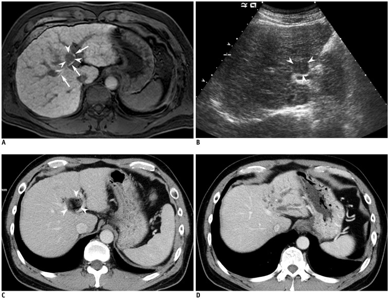
Fig. 1.
1.8 cm sized hepatocellular carcinoma (HCC) in 47-year-old man with liver cirrhosis due to chronic hepatitis B viral infection. He had no previous treatment history for HCC.
A. Axial hepatobiliary phase MRI image (repetition time/echo time, 4.4/2.1 ms) obtained 20 minutes after administration of gadoxetic acid shows 1.8 cm sized HCC lesion in hepatic segment 4, as low signal intensity (arrowheads). Tumor is in contact with hilar bile ducts shown as high signal intensity (arrows). B. On ultrasonogram, tumor in hepatic segment 4 is seen as low echogenecity (arrowheads), in contact with left portal vein at hilar level. Although bile duct is not delineated on this image, it is highly likely that this tumor is contacting left hilar bile duct, as well. C. Axial portal phase CT image obtained immediately after percutaneous ethanol injection (PEI) shows complete ablation of HCC (arrowheads) with no residual tumor. D. Although not shown here, liver CT taken 1 month after PEI demonstrated dilatation of left intrahepatic bile duct. Axial portal phase image of dynamic liver CT obtained 10 months after PEI shows progression of bile duct dilatation, which resulted in atrophy of left hepatic lobe.

