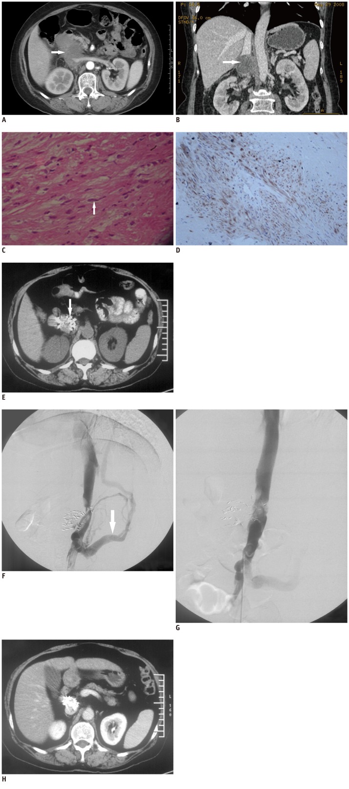
Fig. 1.
Tumor, pathology, treatments and follow-up CT.
Intravenous contrast enhanced CT of mass. Axial view (A, arterial phase) at level of renal vein demonstrates huge mass (white arrow) involving inferior vena cava and partially surrounding left renal vein. Coronal reconstruction (B, venous phase) shows normal corticomedullary enhancement bilaterally, confirming normal function of both kidneys (white arrow). Histology and immunohistochemistry of leiomyo-sarcoma in inferior vena cava. H&E (C, × 200) stain reveals spindle cells (white arrow). Tumor is positive for desmin (D, × 400, brown) indicating leiomyosarcoma deriving from smooth muscle. E. Precontrast follow-up CT three months after two sessions of 125I implantation. Tumor size has markedly decreased and disappeared almost completely. Note high density spots of 125I seeds (white arrow).
Balloon angioplasty for inferior vena cava (IVC) stenosis eight months after 2nd session of 125I seeds implantation. Inferior vena cavography before angioplasty (F) demonstrates stenosis and partial IVC obstruction, with retroperitoneal collateral circulation (white arrow) in keeping with IVC thrombosis. View after cavoplasty (G) shows almost normal lumen of IVC segment. H. Enhanced follow-up CT of 33 months after 2nd session of 125I seeds implantation. CT scan reveals disappearance of tumor without significant caval stenosis or thrombosis.

