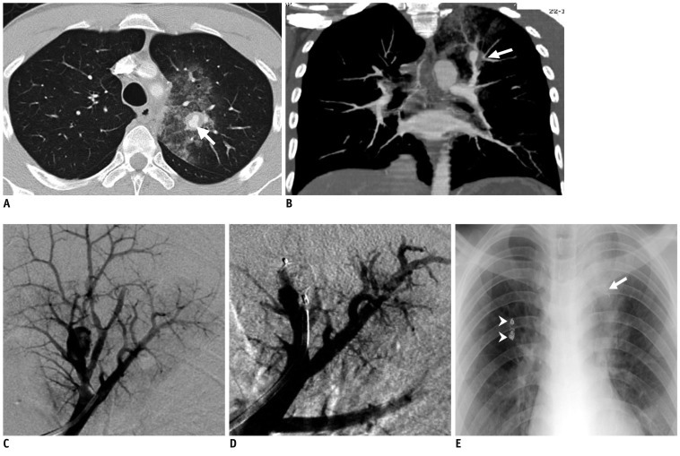
Fig. 1.
Ruptured pulmonary artery aneurysm treated with Ampltazer Vascular Plug 4.
A. Axial contrast-enhanced CT scan (lung window) shows aneurysm of left upper lobe pulmonary artery (arrow) accompanied by mural thrombus and surrounded by diffuse ground-glass opacity at upper left pulmonary lobe, which is suggestive of hemorrhage. B. Coronal maximum-intensity-projection CT image depicts left pulmonary artery aneurysms at level of upper lobe (arrow). C. Left pulmonary angiography via right femoral vein on anteroposterior (AP) projection demonstrates aneurysm at upper lobe pulmonary artery. D. Post-embolization pulmonary angiography on AP projection shows Amplatzer Vascular Plug 4 (AVP 4) with no further filling of aneurysmal sac. E. Chest radiograph demonstrates partial resolution of left upper lobe alveolar-type opacity (still recognizable), presence of AVP 4 (arrow), and coils (arrowheads) due to previous treatment.

