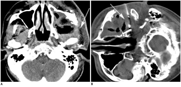
Fig. 1.
Fifty one-year-old male (case 28) with left buccal cancer after surgery and radiotherapy.
A. Contrast-enhanced CT scan shows large necrotic lesion in left masticator space with infiltrative part at periphery (arrow). B. Using subzygomatic approach, 17/18 G biopsy needle set is inserted into lesion (arrow). Biopsy revealed fibrosis with granulation. Follow-up image studies in three and six months showed lesion in stationary status, consistent with biopsy result (not shown).

