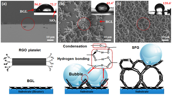
Figure 1.
Schematic illustrations of 3-D SFG formation: (a) BGL: RGO platelets were stacked in the parallel direction with a thickness of 50–100 nm.(b) The structure of SFG seeds formed on the BGL with the help of bubbles via condensation and hydrogen bonding between RGO platelets (denoted as red circles and boxes). and (c) SFG formation from the seed and bubbles.

