
Figure 2.
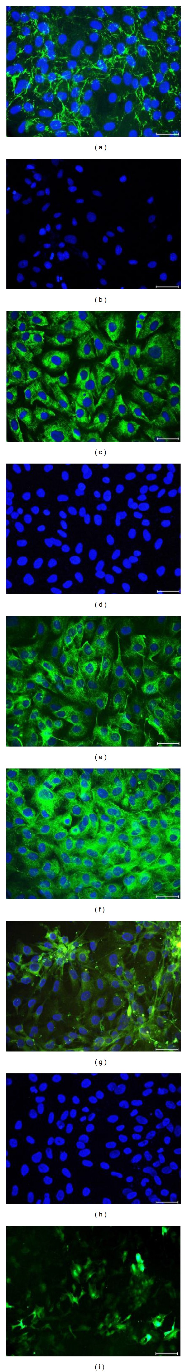
Immunofluorescence staining of rAT-MSCs for fibronectin (a), CD34 (b), GFAP (c), CD45 (d), Nestin (e), Vimentin (f), Map2a, b (g), and Cytokeratin 18 (h). Staining pattern was cytoplasmic for fibronectin, GFAP, nestin, and vimentin; and both membranous and cytoplasmic for Map2a, b. After transfection, rAT-MSCs showed GFP+ immunostaining (i). Nuclei were labeled with DAPI (blue). Scale bars: 50 μm.