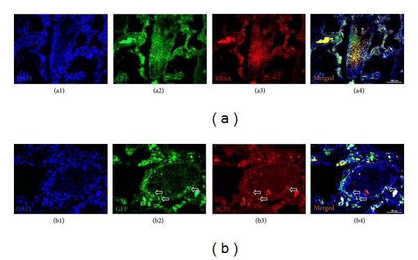
Figure 6.

Immunostaining of GFP+/VASA+ and GFP+/SCP1+ cells in busulfan-treated testis with rAT-MSCs injection. GFP+/VASA+ cells were localized in the seminiferous tubules of testis (white star), but most of the tubules were empty (blue asterisk) (a1–a4). The expression of meiosis marker SCP1 in GFP+ cells might indicate the transdifferentiation of MSCs into spermatogenic cells (white arrows) (b1–b4).
