
Figure 7.
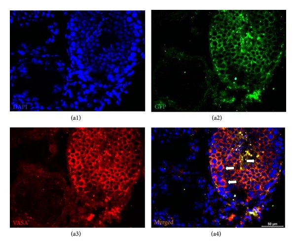
GFP+ cells in seminiferous tubule. MSC-received testis was fixed and stained for GFP (green) and spermatogenic cell marker, VASA (red). The cells in seminiferous tubule with dual staining were shown by arrows (a4). The adjacent tubules showed no staining for VASA, which indicated the absence of spermatogenic activity.