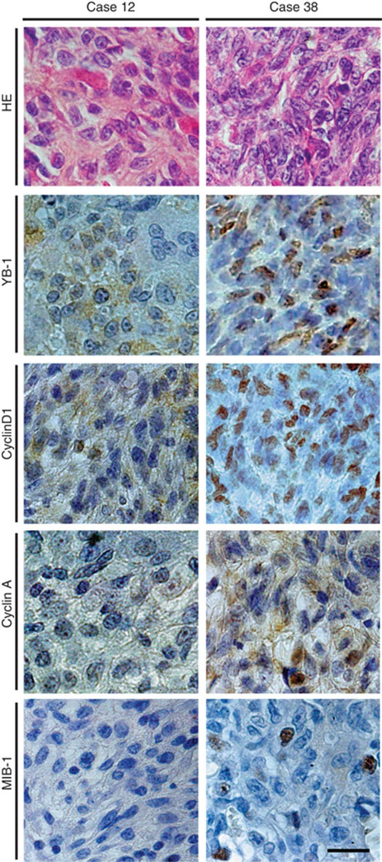
Figure 8.
Haematoxylin−eosin and immunohistochemical staining of human OS sections. Representative staining of YB-1, cyclin D1, cyclin A, and MIB-1 in OS samples. Paraffin sections were stained with haematoxylin−eosin and immunohistochemically stained using anti-YB-1, anti-cyclin D1, anti-cyclin A, and anti-YB-1 antibodies, then were visualised using the diaminobenzidene substrate system. Counterstaining was then performed using diluted haematoxylin. In case 38 (YB-1 nuclear expression positive, died of disease), high levels of cyclin D1 (⩾10%) and cyclin A (⩾40%) expression were evident, whereas in case 12 (YB-1 nuclear expression negative, continuously disease free), expression of cyclin D1 and cyclin A were low. Scale bar, 20 μm.

