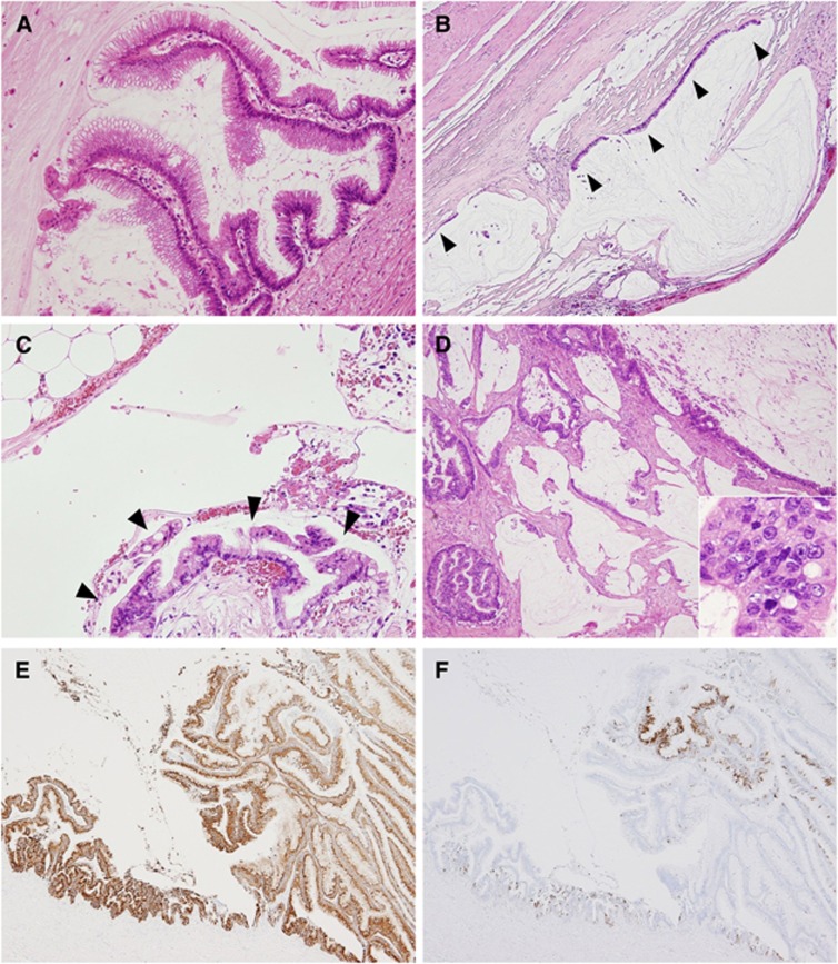
Figure 1.
Histology of appendiceal mucinous tumours. (A–E) Histological and immunohistochemical findings of LAMN (A–C, E, F) and MAC (D). LAMN exhibiting a villous growth pattern (A). The tumour cells had low-grade cytological atypia. Mucin pools were present in the subserosal layer and were partly lined by mucinous epithelium (arrowheads). The stroma showed hyalinizing fibrosis (B). Peritoneal deposits of LAMN (C). Low-grade tumour cells grew on the surface of the omentum without evidence of stromal invasion (arrowheads) (C). MAC. Abundant mucin production and a frankly invasive growth pattern were evident. The tumour cells showed cytologically high-grade atypia, with a high nuclear–cytoplasmic ratio and prominent nucleoli (inset) (D). LAMN exhibiting diffuse MUC2 expression (E) and focal MUC5AC expression (F).

