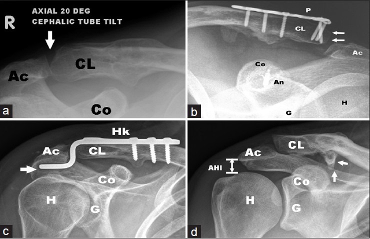
Figure 2.

Radiographic outcomes of surgical treatment at follow-up are shown. (a) Acromioclavicular joint degeneration (arrow). (b) Acromioclavicular joint subluxation (arrows), (c) Hook migration and osteolysis of acromial undersurface (arrow), (d) Peri-coracoid ossification (arrows). (Ac: acromion, CL: clavicle, Co: coracoid, P: plate, An: suture anchor, G: glenoid, H: humeral head, Hk: hook plate, AHI: acromiohumeral interval)
