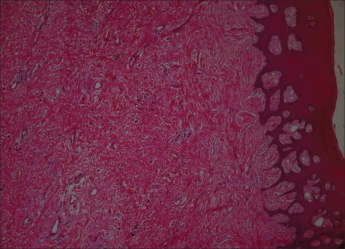
Figure 2.

Stained section shows relatively thick parakeratinized epithelium with underlying connective tissue composed of interlacing thick bundles of collagen fibres, fibroblasts, few blood vessels and areas of chronic inflammatory cell infiltrate (H and E, 4×)
