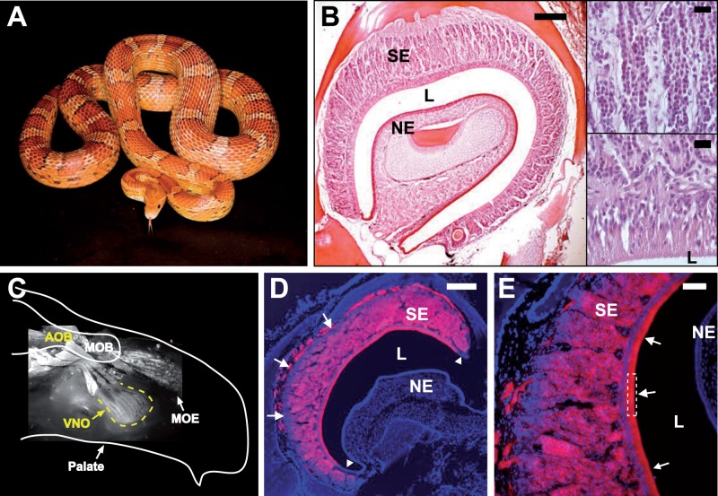
Fig. 1.—
The VNO of the corn snake. (A) Corn snake. (B) Coronal section of a corn snake VNO stained with hematoxylin and eosin (HE). Full organ (left), scale bar 200 μm; close-ups of the columnar sensory epithelium (top right) and the zone in contact with the outside world (bottom right), scale bars 20 μm; SE, sensory epithelium; NE, nonsensory epithelium; L, lumen. (C) Schematic representation of a head hemi-section with a picture of the fluorescence detected in the VNO and the main olfactory epithelium (MOE) after application of a retrograde tracing dye onto the AOB and main olfactory bulb (MOB). (D,E) Coronal section of the VNO with fluorescence (red) detected after application of the tracing dye onto the AOB. Arrowheads and arrows in (D) indicate groups of potentially immature neurons at the edges and base, respectively, of the neuroepithelium; arrows and dotted frame in (E) indicate dendritic projections to the lumen. DNA is stained with Hoechst (blue). Scale bar in (D), 200 μm and in (E), 50 μm.

