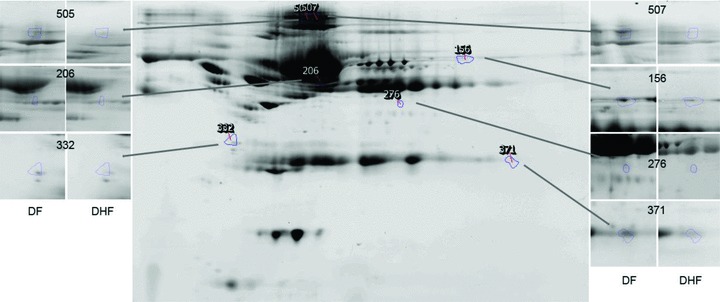
Figure 3.

Shown is a reference gel of 2‐DE of BAP fractionated and IgY depleted plasma from the study subjects. The location of protein spots that contribute to the prediction of DHF are indicated. Insets, spot appearances for reference gels for DHF and DF. Spot 156 (C4A), 206 (albumin * 1), 276 (fibrinogen), 332 (tropomyosin), 371 (immunoglobulin gamma‐variable region), 506 (albumin * 2), and 507(albumin * 3).
