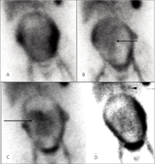
Figure 1. Selected images from the one minute per frame sequentialimages of an in vitro gastrointestinal bleeding scan on a 6-month-oldpregnant woman. At 4 minutes after injection (A) there is traceraccumulation in the liver, spleen, the major blood vessels and theuterus. Increased tracer accumulation is seen in lateral walls of theuterus more on the left than the right, during the beginning of thestudy. This likely represents the normal perfusion to the placenta. At15 minutes, time there is diffuse activity seen over the gestationaluterus. Faint area of mildly increased activity accumulation within theuterus is noted. This central activity changes position over the courseof the scan represents the viable fetus (B). The activity in the fetus isprobably due to radioactive break down components of Tc-99m RBCssince intact RBCs do not normally cross the placenta. At 40 minutes(C) there is seen to be more prominent activity accumulation in theviable fetus. The final image at 60 minutes (D) shows a more clearlydefined “blush” of activity in the left upper quadrant questionablefor a GI bleeding (arrowhead) and an angiography showed a gastrosplenicarteriovenous malformation (AVM).

