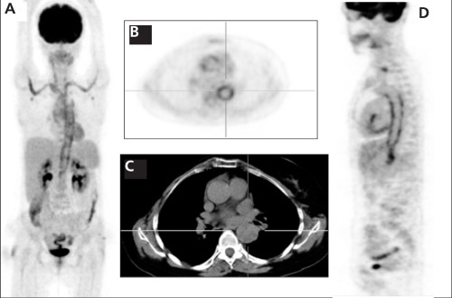
Figure 1. A 64-year-old woman with active Takayasu arteritis. Figure1A (MIP), Figure 1B (Axial PET), Figure 1D (Sagittal PET) imagesdemonstrated intense F 18 FDG uptake in the aorta, bilateral subclavianand brachiocephalic arteries consistent with Takayasu arteritis. The maximum standardized uptake value of the aorta was 9.2 beforetherapy.

