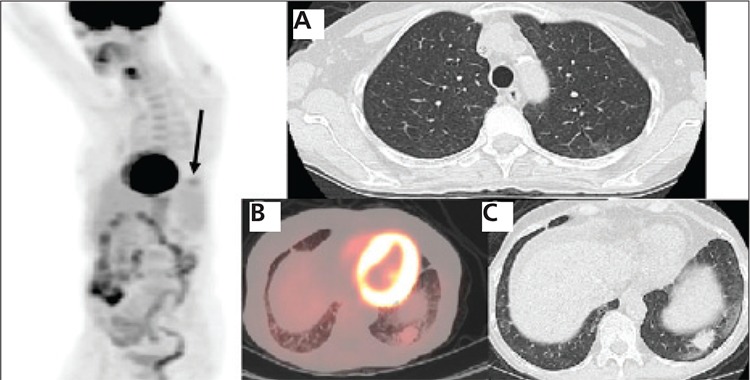
Figure 3. Two months after Rituxan therapy a restagingPET/CT shows a faint focus of abnormal increased FDG uptake inthe left lower lateral chest (arrow) and complete resolution of theFDG uptake in the upper medial chest on the 3D PET image. On theaxial CT image (A) the upper lobe lesion has completely resolved. Thefused axial image (B) and axial CT slice (C) show interval decrease inthe size and FDG uptake of the lower lobe lesion, to 2.4x1.7 cm withSUV max of 2.4, representing favorable response to treatment.

