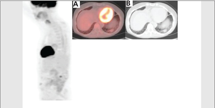
Figure 4. Six months after completion of therapy, 3DPET image shows interval near complete resolution of FDG uptake inthe left lower lateral chest. The fused axial image (A) and axial CTslice (B) demonstrate decrease in the size of left lower lobe lesionwhich has only subtle increased FDG uptake, the lesion at this timemeasured 2.4x1.4 cm with SUV max of 1.7, representing continuedfavorable response to treatment.