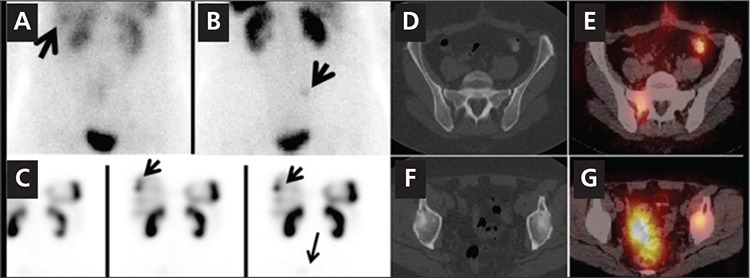
Figure 3. A 58 year-old woman with carcinoid tumor of the small bowel withknown liver metastases had an In-111 Pentetreotide scan. Anterior (A) andposterior (B) planar images of the abdominopelvic region show abnormal focaluptake is noted in the inferior right lobe of the liver (arrow, image A) and afaint focus of increased uptake is noted in the mid upper right hemi pelvis(arrow, image B). Selected coronal SPECT (C) images show foci of increaseduptake in the liver, corresponding to the known sites of liver metastases (smallarrows). A very faint focus of tracer uptake slightly above the bladder on theright is suspicious for a metastasis (arrow, C). However accurate interpretationof this finding is not possible with SPECT scan only. Axial SPECT/CT images atthe level of pelvis (D, E, F, G) show increased tracer uptake in the right sacrum(D,E), consistent with early bone metastasis, which correlated to the suspiciousfinding on planar posterior image (B). Another focus of abnormal increasedtracer uptake was noted in left acetabulum with predominantly scleroticchanges in the acetabulum (F, G). This lesion was not visible on planar imagesand was not localizable on SPECT alone. SPECT /CT however detected andlocalized this metastatic bone lesion.