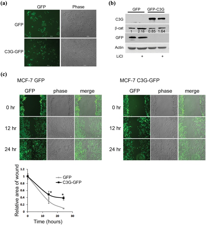
Figure 6.
Stable expression of C3G inhibits motility of MCF-7 cells. (a) Live cell images of MCF-7 clones stably expressing GFP or C3G-GFP. Bar, 50 µm. (b) Endogenous β-catenin expression in MCF-7 clones. MCF-7 clones stably expressing GFP or C3G-GFP were either left untreated or treated with LiCl and lysed after 24 hours of treatment. Cell lysates were subject to Western blotting to detect the indicated proteins with antibodies. (c) Effect of C3G expression on cell motility. Wound healing assay was performed on MCF-7 clones stably expressing GFP or C3G-GFP. Live cell images were taken after indicated time periods of making the wound. Graph shows relative wound area with respect to time averaged from 3 wounds each from 2 independent experiments. Student t test was performed to test the significance of difference in wound area. Bar, 50 µm. *P < 0.05.

