Figure 7.
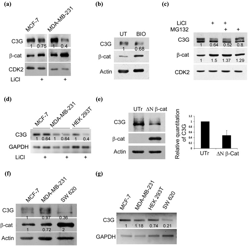
β-catenin activation reduces cellular C3G levels. (a) Increase in cellular β-catenin levels by GSK3β inhibition results in reduced C3G levels. C3G and β-catenin levels determined by Western blotting in MCF-7 and MDA-MB-231 cells treated with LiCl. CDK2 was used as a loading control. (b) C3G and β-catenin levels in MCF-7 cells treated with BIO, a specific inhibitor of GSK3β. UT, untreated. (c) β-catenin activation induces reduction in C3G levels independent of proteasomal degradation. C3G and β-catenin levels were examined under conditions of LiCl and MG132 treatment in MCF-7 cells. CDK2 was used as internal control. (d) Activation of β-catenin by LiCl treatment reduces C3G transcript levels. MCF-7, MDA-MB-231, and HEK 293T cells were treated with 50 mM LiCl, and cells were lysed after 24 hours for RNA isolation. cDNA was prepared from RNA followed by semiquantitative PCR using primers designed for GAPDH and C3G. (e) Expression of a constitutively active variant of β-catenin reduces cellular β-catenin protein levels. Cell lysates of HEK 293T cells transfected with ΔN β-catenin for 48 hours were subject to Western blotting to detect C3G and β-catenin expression. Actin was used as a loading control. UTr, untransfected. Bar diagram shows relative C3G levels as mean ± SD from 3 experiments. (f) Comparative C3G expression in cell lines. Cell lysates of MCF-7, MDA-MB-231, and SW620 cells were subject to Western blotting to detect the indicated proteins with antibodies. SW620 cells show constitutively high β-catenin activity due to APC mutation. (g) Semiquantitative PCR to detect C3G and GAPDH transcripts in MCF-7, MDA-MB-231, HEK 293T, and SW620 cells.

