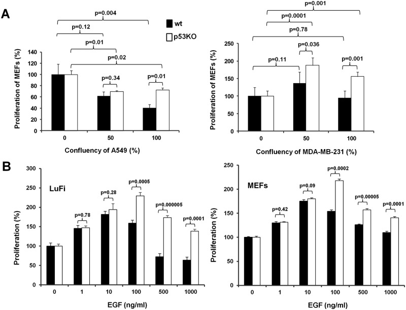
Figure 1.
Proliferation of primary mouse fibroblasts in conditioned media from cancer cells and at increasing concentrations of EGF. (A) Proliferation of MEFs differing in p53 status in conditioned media from A549 lung (left) or MDA-MB-231 breast (right) human cancer cells grown under semiconfluent or confluent conditions. The P values of various pairwise comparisons are also indicated in the figure. (B) Proliferation of LuFi (left) or MEFs (right) differing in the status of p53 at increasing concentrations of EGF. The concentration of EGF is indicated.

