
Figure 2.
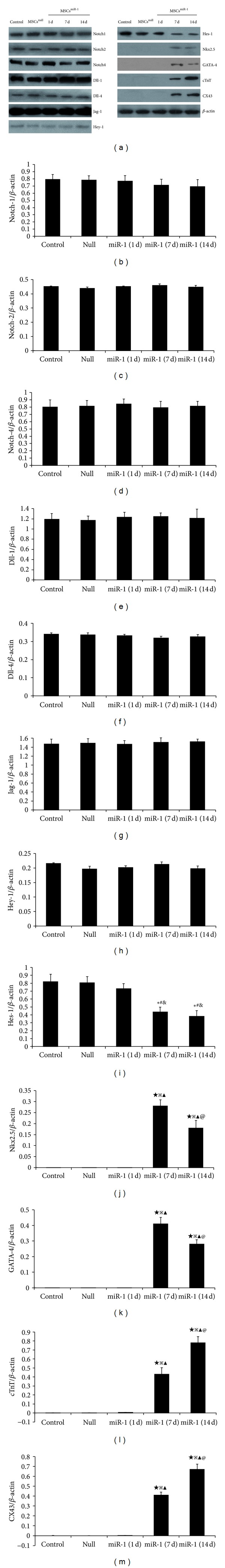
Western blot was performed for Notch signaling and cardiomyocyte-specific markers in MSCs, MSCsnull, and MSCsmiR-1 (1 d, 7 d, and 14 d). (a) Expression of Notch-1, Notch-2, Notch-4, Dll-1, Dll-4, Jag-1, Hes-1, and Hey-1 were detected on MSCs. Semiquantitative data showed that the ratio of optical density for Notch-1, Notch-2, Notch-4, Dll-1, Dll-4, Jag-1, and Hey-1 did not alter in MSCsmiR-1 on days 1, 7, and 14 (b)–(h). The expression of Hes-1 (i) in MSCsmiR-1 was decreased by days 7 and 14. In MSCsmiR-1, the expression of Nkx2.5 (j) and GATA-4 (k) were detected on day 7 and decreased by day 14. cTnT (l) and CX43 (m) expression were detected on day 7 and significantly increased by day 14 (control = MSCs; null = MSCsnull = MSCs infected with mock lentiviral vectors without miR-1; miR-1= MSCsmiR-1 = MSCs infected with miR-1 recombinant lentiviral vectors; compared to MSCs, *P < 0.05, ★ P < 0.01; compared to MSCsnull, # P < 0.05, ※ P < 0.01; compared to MSCsmiR-1 (1 d), & P < 0.05, ▲ P < 0.01; compared to MSCsmiR-1 (7 d), @ P < 0.05).