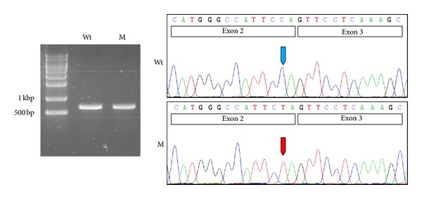
Figure 2.

In vitro splicing assay. RT-PCR analyses of the wildtype (Wt) and mutated (M) pSPL3 vector. Both showed one normally spliced product that was confirmed by sequencing. The sizes of the marker band are shown in the gel image. Exons are shown in the box in the sequencing chromatogram. Wildtype and mutated nucleotide are indicated by blue and red arrows, respectively.
